Application
RNA Sequencing
Explore Gene Expression at Scale
RNA sequencing from bulk tissues to single cells, revealing transcriptional regulation, alternative splicing, and cellular heterogeneity.

Application
Explore Gene Expression at Scale
RNA sequencing from bulk tissues to single cells, revealing transcriptional regulation, alternative splicing, and cellular heterogeneity.
NGS-based RNA sequencing (RNA-seq) converts RNA into cDNA fragments through reverse transcription, followed by high-throughput short-read sequencing. Without relying on predefined microarray probes, RNA-seq provides unbiased digital quantification, enabling accurate measurement of gene expression abundance across an ultra-wide dynamic range.
Quantify differential gene expression across tissues, developmental stages, and drug-induced conditions.
Identify novel transcripts, alternative splicing events, and gene fusions.
Characterize disease-associated regulatory networks and non-coding RNA biomarkers in cancer and complex diseases.
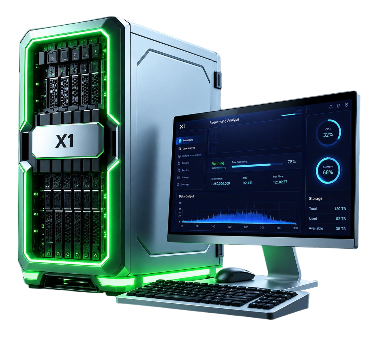
X1 Bioinformatics Workstation is an integrated bioinformatics solution, designed to streamline sequencing data analysis from raw data to application-ready reports. Featuring flexible hardware configurations and pre-built bioinformatics pipelines, it delivers plug-and-run, end-to-end data analysis for diverse laboratory throughput needs.
X1 requires no complicated deployment or professional bioinformatics setup, allowing one-click standardized analysis to cut hands-on work, shorten turnaround time, boost data security and deliver actionable sequencing results faster.
Supports multiple bioinformatics plugins for a wide range of analysis applications.

Runs reliably offline to help keep sensitive data secure and private.

No complex command-line operations required, enabling streamlined analysis with minimal training.

Real-time memory and storage monitoring with early warnings to help prevent capacity issues.
01
Bulk RNA-seq enables transcriptome-wide profiling through mRNA enrichment or rRNA depletion. mRNA sequencing targets protein-coding transcripts, while Total RNA sequencing retains both mRNA and long non-coding RNA (lncRNA) following rRNA depletion.
Extract total RNA and perform stringent RNA integrity assessment, including RIN evaluation.
Use magnetic beads to capture poly(A)-tailed mRNA, or enzymatic methods or hybridization probes to directly remove ribosomal RNA.
Randomly fragment RNA and use random primers for reverse transcription to synthesize double-stranded cDNA.
Perform end repair, A-tailing, paired-end adapter ligation, and PCR amplification, followed by paired-end sequencing.

Detect gene expression changes with high sensitivity, including low-abundance transcripts.
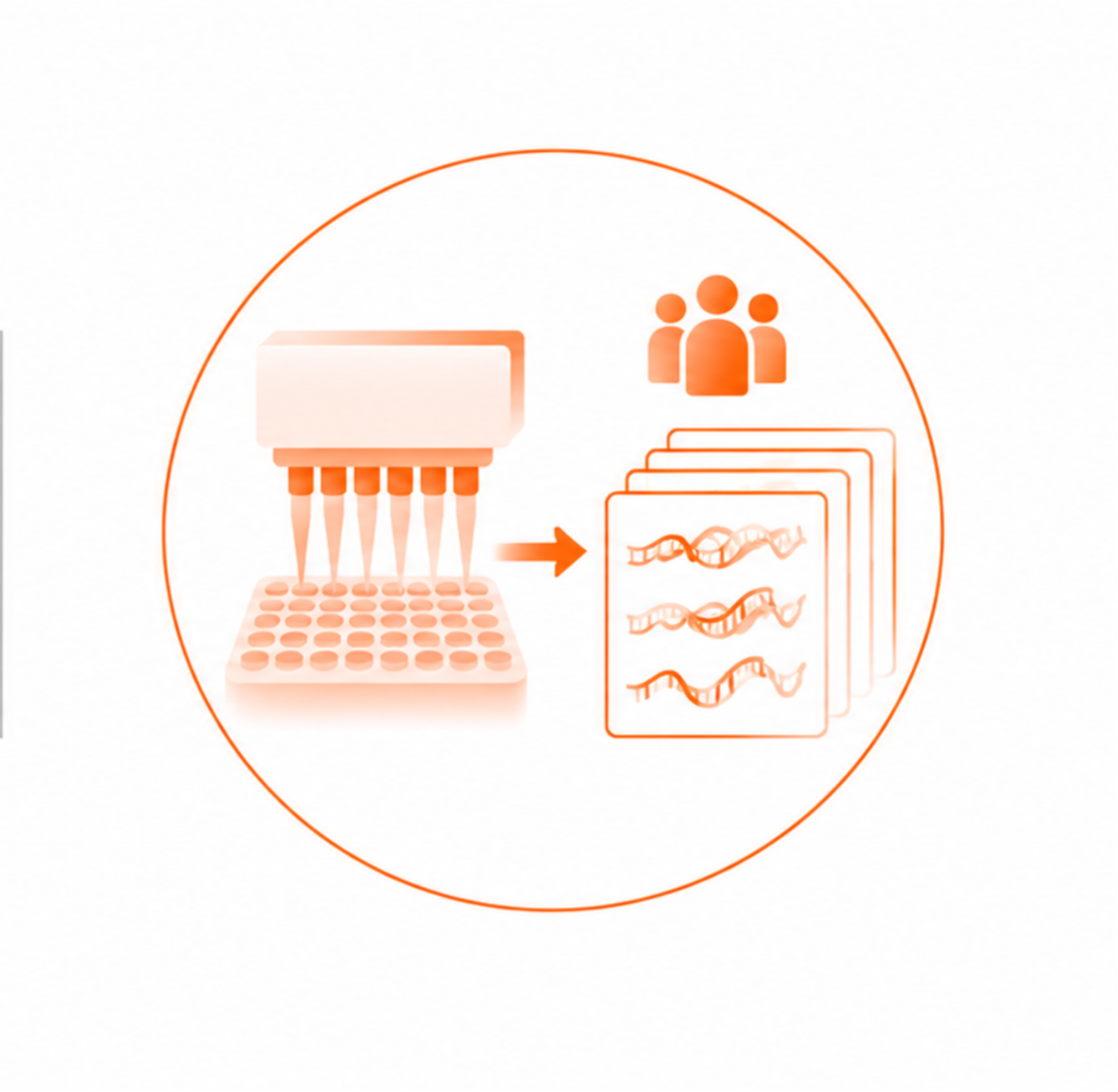
Automated library preparation supports efficient expression profiling across large clinical and research cohorts.
02
Small RNA sequencing enriches and quantifies short non-coding RNAs of approximately 18–30 nt, including miRNAs, siRNAs, and piRNAs. These small RNAs play key roles in post-transcriptional regulation.
Use polyacrylamide gel electrophoresis (PAGE) or specialized magnetic beads to selectively isolate short RNA fragments of approximately 18–30 nt.
Directly ligate single-stranded RNA adapters to the 3′ and 5′ ends of small RNA molecules without fragmentation.
Reverse-transcribe the RNA into cDNA followed by PCR amplification.
Due to the short fragment length, single-end sequencing, such as SE50 or SE75, is typically used for efficient sequencing.
Preserves native full-length sequences and terminal information without fragmentation, enabling high-resolution analysis.
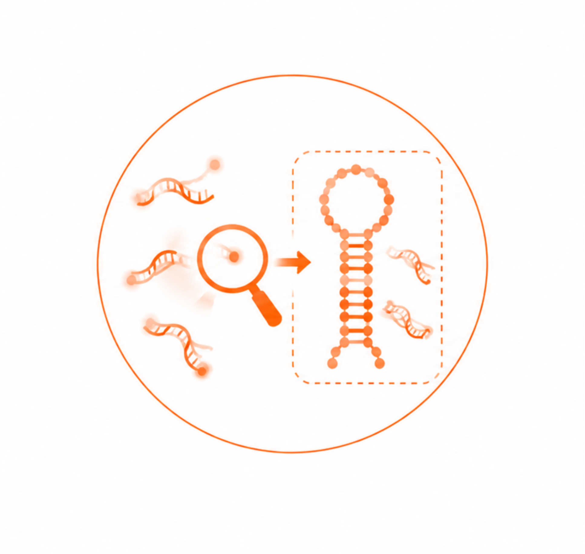
Enables de novo identification of novel small RNAs and their precursor structures without prior sequence information.
03
Single-cell RNA sequencing resolves cellular heterogeneity beyond bulk RNA-seq. Microfluidic or microwell technologies enable transcriptome profiling of thousands of individual cells at single-cell resolution.
Dissociate tissues into a high-viability single-cell suspension.
Use a microfluidic system, such as the 10x Genomics platform, to encapsulate individual cells together with gel beads carrying cell barcodes and unique molecular identifiers (UMIs) within droplets.
Lyse cells within droplets. Released mRNA molecules are labeled with cell-specific barcodes to identify their cell of origin and UMIs to distinguish individual transcript molecules.
Break the droplets, pool cDNA from all cells, and perform high-depth paired-end NGS.
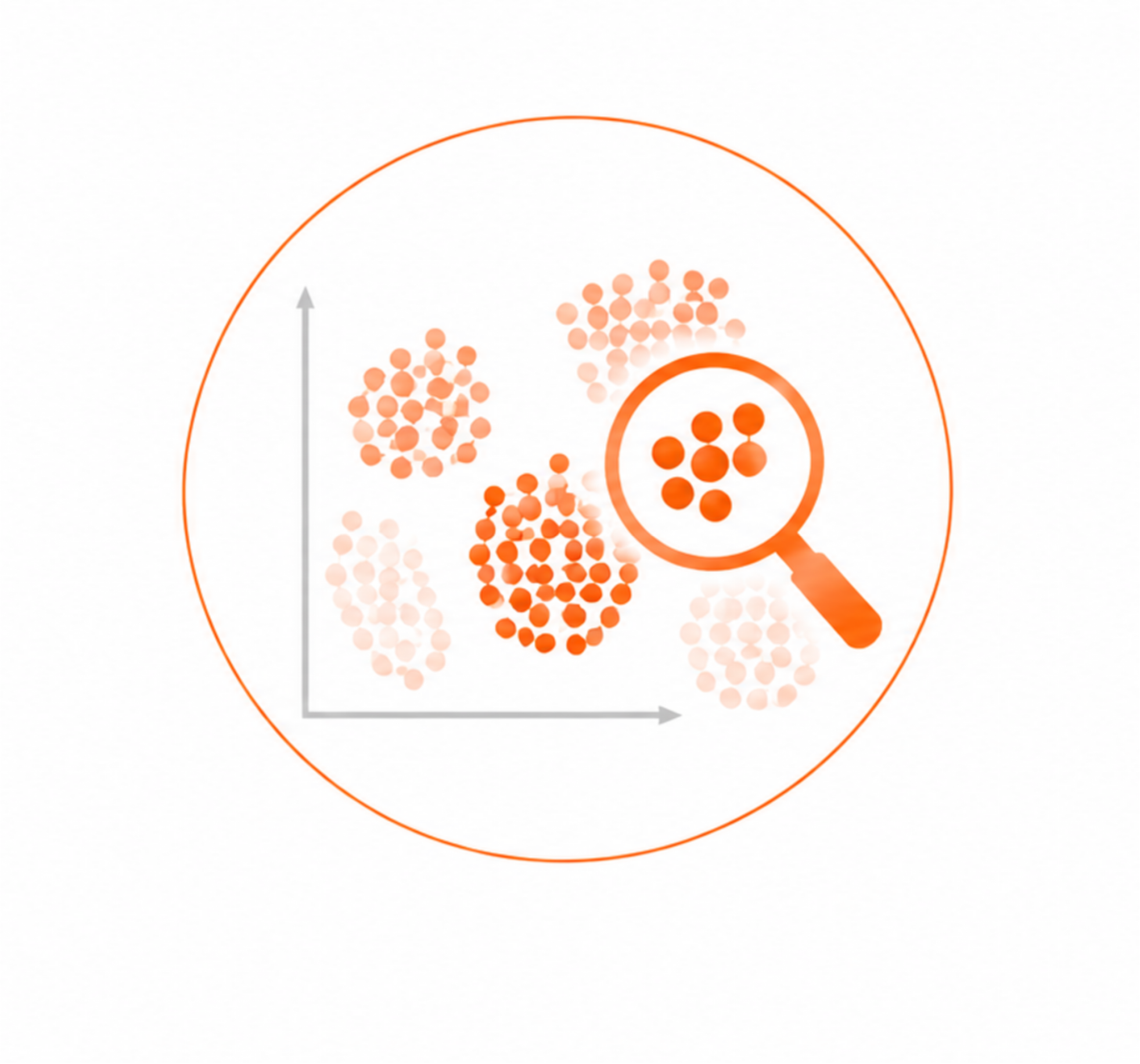
Identify rare cell types and define novel cell subpopulations.
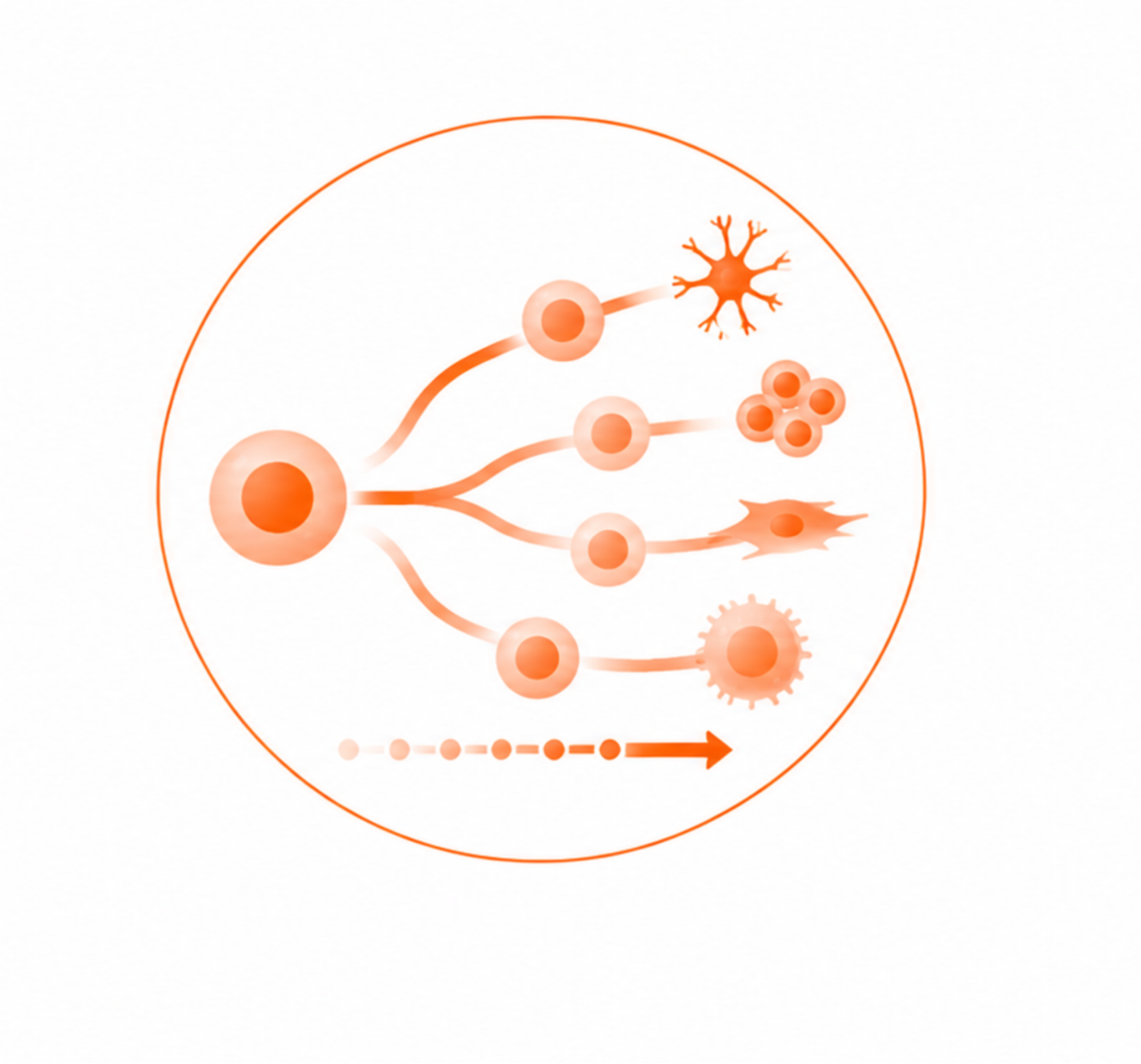
Use bioinformatics algorithms to infer pseudotime trajectories of cellular differentiation and development, providing insights into dynamic cellular changes.
04
Spatial transcriptomics maps gene expression within the native tissue context. Spatially barcoded arrays enable high-throughput transcriptome profiling while preserving the spatial organization of cells.
Place fresh-frozen or FFPE tissue sections onto specialized chips containing spatial barcode arrays.
Perform H&E or immunofluorescence staining followed by microscopic imaging to record tissue morphology.
Permeabilize the tissue to release mRNA, which is captured in situ by spatially barcoded primers on the chip surface.
Recover spatially barcoded cDNA, construct sequencing libraries, and perform NGS. Integrate sequencing data with microscopy images based on spatial coordinates.
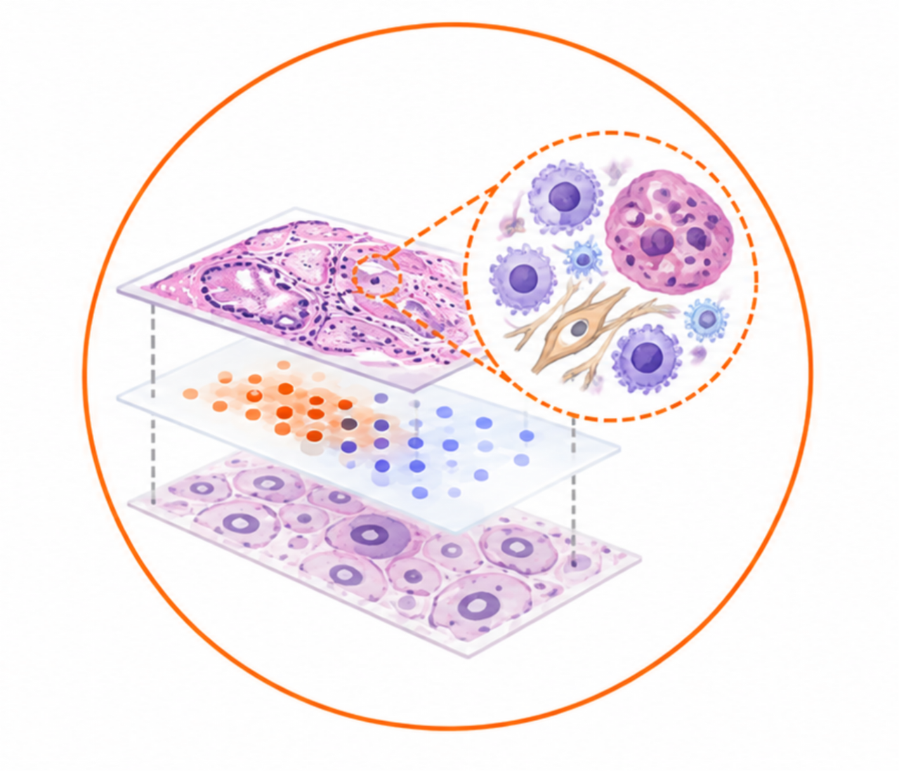
Preserving complex tissue architecture without tissue dissociation, including the tumor microenvironment (TME), neuroanatomical structures, and neighboring cell-cell interactions.
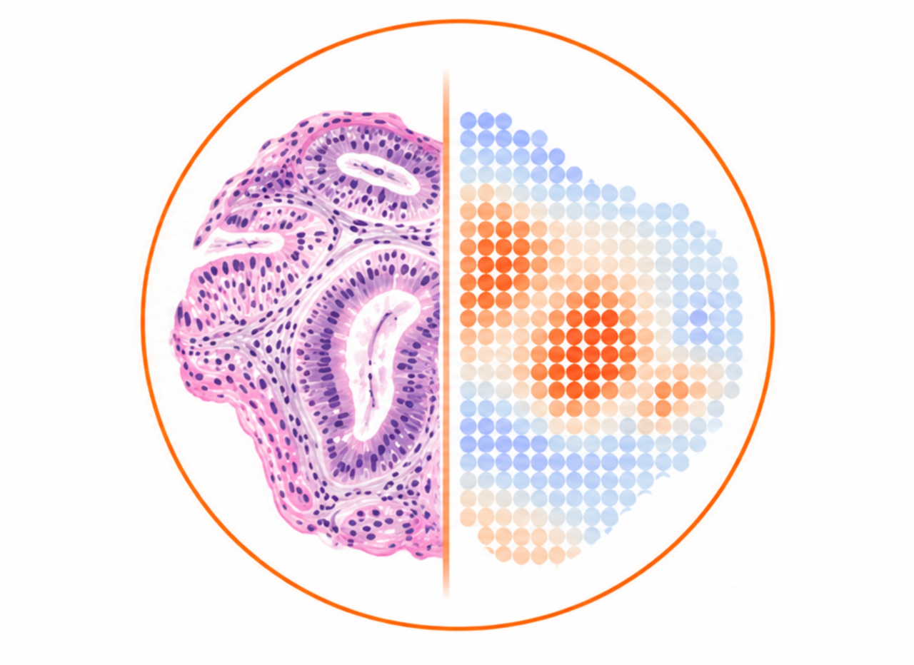
Integrating conventional histopathological visualization with high-throughput quantitative transcriptomic profiling.